|
|
Research Methods
| ★Method 1 ― OCT image segmentation using graph based methods |
| We have reported a graph-theoretic based method for abnormal retinal layer segmentation and SEAD segmentation. The proposed graph-theoretic based method effectively combined the GS and GC methods for segmenting the retinal layers and the SEADs simultaneously. |
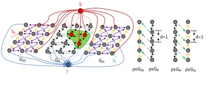
Graph construction of graph-search-graph-cut method
|
Segmentation of fluid-associated abnormalities in Retinal OCT
|

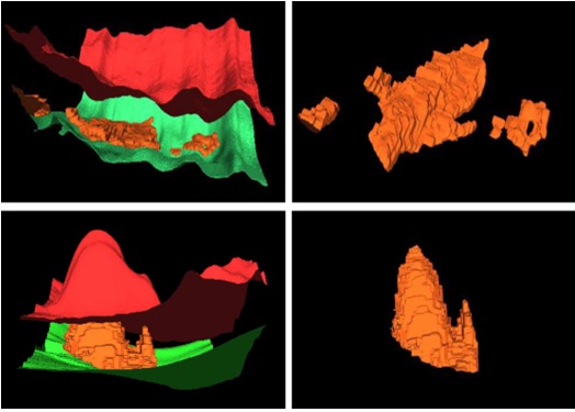
Segmentation of OCT images with Serous Pigment Epithelial Detachments |
Related papers |
(1). Xinjian Chen, Meindert Niemeijer, Li Zhang, Kyungmoo Lee, Michael D. Abràmoff, and Milan Sonka, 3D SEAD Segmentation by Probability Constrained Graph-Search-Graph-Cut, IEEE Transactions on Medical Imaging, Vol. 31, No.8, pp.1521-1531,Epub. Mar. 19, 2012.
(2). Fei Shi, Xinjian Chen*, Heming Zhao, Weifang Zhu, Dehui Xiang, Enting Gao, Milan Sonka and Haoyu Chen, Automated 3-D Retinal Layer Segmentation of Macular Optical Coherence Tomography Images with Serous Pigment Epithelial Detachments, IEEE Transactions on Medical Imaging, 2014.
|
| ★Method 2 ― Abnormal detection using classification methods |
| Classification is a common research field in machine vision and pattern recognition, which has been applied for medical image detection, medical image segmentation and medical diseases diagnosing. Classification involves two-class and multi-class classification. |
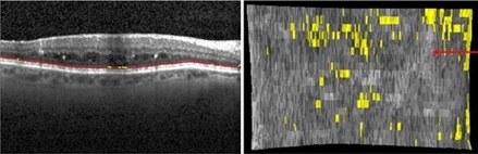
Quantification of External Limiting Membrane Disruption
|
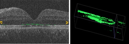
Quantification of IS/OS junction Disruption
|
Related papers |
(1). Xinjian Chen, Ping Hou, Chao Jin, Weifang Zhu, Xiaohong Luo, Fei Shi, Milan Sonka and Haoyu Chen, Quantitative analysis of retinal layers' optical intensities on 3D optical coherence tomography, IOVS, vol. 54, No. 10, pp. 6846-6851,Oct, 2013.
(2). Xinjian Chen, Li Zhang, Elliott H. Sohn, Kyungmoo Lee, Meindert Niemeijer, John Chen, Milan Sonka, and Michael D. Abramoff, Quantification of External Limiting Membrane Disruption Caused by Diabetic Macular Edema from SD-OCT, IOVS, Vol. 53, No. 13, pp. 8042-8048, 2012.
|
| ★Method 3 ― 3D Shape Constrained Graph Cut |
| The executable version of 3D shape constrained GC with user interface can be downloaded (please read the instructions from http://www.mipg.upenn.edu/~cavass/ before downloading. This is a zip file, delete extention “.odt” after downloading. ). GC user interface with CAVASS, more infomation about cavass can be found from official website: http://www.mipg.upenn.edu/~cavass/ |
|
  |
|
| The 3D Shape Constrained GC |
| ★Method 4 ― Graph Cut and Oriented Active Appearance Models |
| we have proposed a synergistic combination of the image-based GC and model-based ASM methods. The proposed cost function effectively integrates the ASM shape information into the GC paradigm. The automatic object recognition strategy employed by GC-ASM is based on the minimum total GC cut cost, which utilizes combined prior shape and specific image information. |
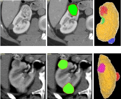
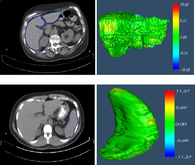
Tumor growth prediction
|
Multiple organ segmentation |
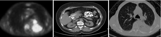
Lung tumor segmentation on PET-CT image |
Related papers |
(1). GC-ASM: Synergistic Integration of Graph-Cut and Active Shape Model Strategies for Image Segmentation, Computer Vision and Image Understanding, 2013.
(2). Xinjian Chen, et. al, Medical Image Segmentation by Combining Graph Cut and Oriented Active Appearance Models, IEEE Transactions on Image Processing, Vol.21, No.4,pp. 2035-2046, EPub. Jan. 31,2011.
(3). Kidney Tumor Growth Prediction by Coupling Reaction-Diffusion and Biomechanical Model, IEEE Transactions on Biomedical Engineering, 2013.
(4). Xinjian Chen, Jayaram K. Udupa, Abass Alavi and Drew A. Torigian, Automatic Anatomy Recognition via MultiObject Oriented Active Shape Models, Medical Physics, Vol. 37, No. 12, pp. 6390-6401, 2010.
|
| ★Method 5 ― Phase-resolved Doppler OCT |
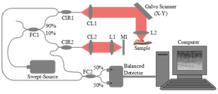
Principle of Phase-resolved Doppler OCT
|
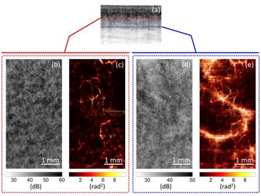
In Vivo Doppler OCT of Human Palm |
| ★Method 6 ― Enhanced low-rank + sparsity decomposition for speckle reduction in optical coherence tomography |
Speckle artifact can strongly hamper quantitative analysis of optical coherence tomography (OCT). We introduce a new method for speckle reduction, which leverages from low-rank + sparsity decomposition (LRpSD) of OCT images, we combine nonconvex regularizaiton-based low-rank approximation of original OCT image with sparsity term that incorporates the speckle. The method yields OCT image with improved contrast-to-noise ratio, contrast and edge fidelity. [Matlab codes for Download]
This work is funded by the 2015-2017 bilateral Chinese-Croatian grant "Nonlinear sparse component analysis approach to decomposition and contrast enhancement of PET/CT and OCT images".
|

|
Related papers |
Ivica Kopriva, Fei Shi, Xinjian Chen, Enhanced low-rank +sparsity decomposition for speckle reduction in optical coherence tomography, Journal of Biomedical Optics, 2016 (In Press)
|
|